What is a scleral buckle?
What is a scleral buckle?
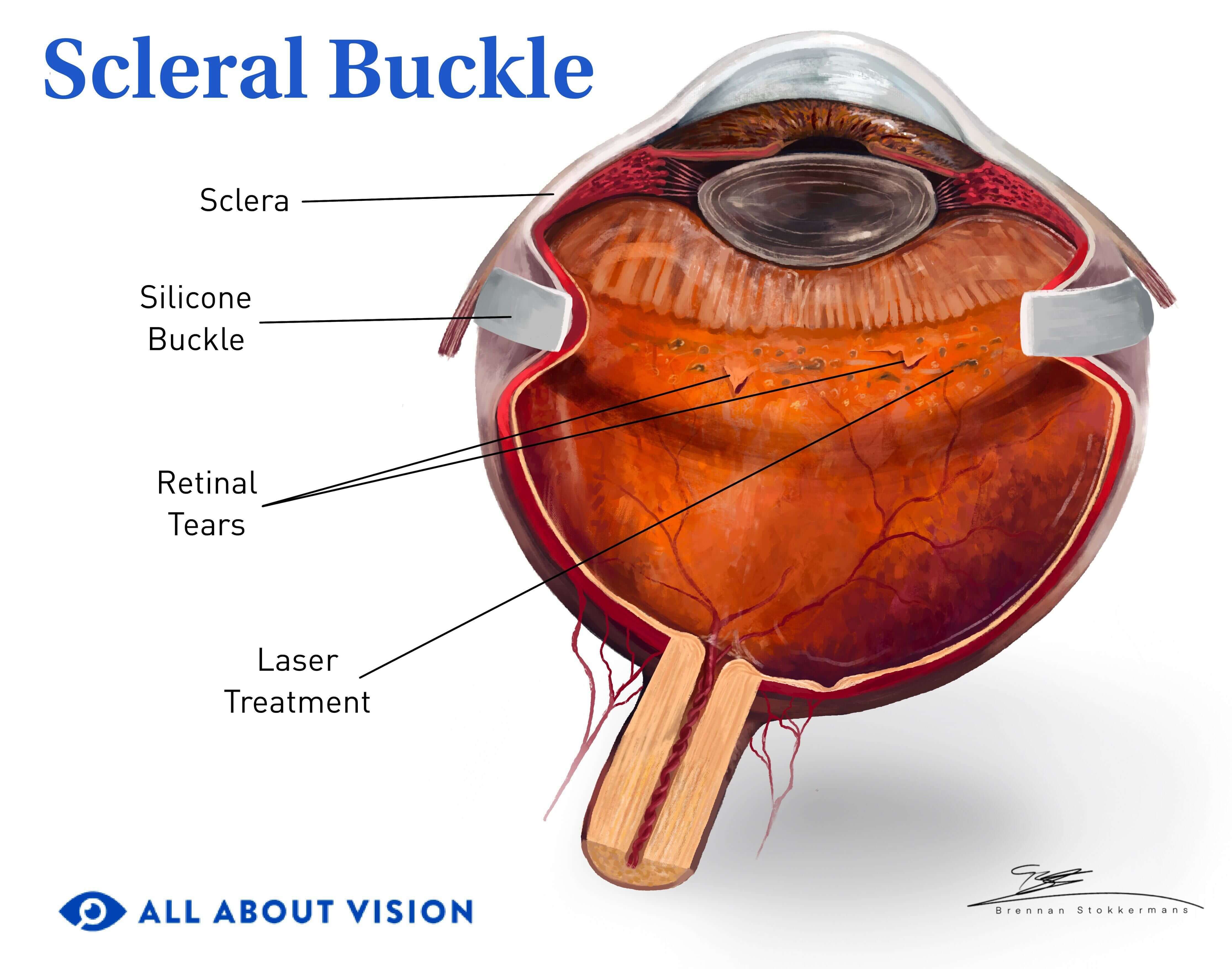
A scleral buckle is a surgery that places a permanent silicone band around the eye to help return the retina — a thin layer of tissue located in the back of the eye — into position after a retinal detachment. It wraps around the eyeball like a belt, supporting the retina.
The retina successfully reattaches in about 90% of patients who have scleral buckle surgery. It is performed in an operating room and can require several weeks to fully recover. Some discomfort may be present the first few days after the surgery, but pain medication will be prescribed to help with discomfort.
Scleral buckle surgery may be performed in combination with other procedures to repair a retinal detachment. This includes vitrectomy, which removes and replaces the gel-like substance inside the eye to decrease traction on the retina. Cryopexy (freezing) may also be performed during scleral buckle surgery to help seal retinal holes or tears and reattach the retina.
Ophthalmologists have successfully performed this eye surgery for over 50 years. As new procedures have emerged, it is not as common as it used to be. However, it is still considered an important treatment option for some cases of retinal detachment.
Why may I need scleral buckling?
Scleral buckling is a treatment eye surgeons sometimes use to repair a detached retina. When the retina detaches, it can lead to profound vision loss. The goal of repairing a detached retina is to recover as much vision as possible.
If someone is experiencing symptoms that indicate a possible retinal detachment, an eye doctor will perform a comprehensive eye exam. If a detachment is detected, they will recommend the appropriate procedure to reattach the retina. The treatment will depend on the severity of the retinal detachment and the patient’s symptoms.
Some procedures can be done in the office, while other treatments, such as scleral buckling or vitrectomy, require an operating room. If a patient is not having symptoms and the detachment is small, the doctor may treat with a laser to help reattach the retina. Another in-office option is pneumatic retinopexy, the injection of a gas bubble to provide light pressure against the retina.
When the retinal detachment is more extensive, a scleral buckle may be needed to support the retina from outside the eyeball. Once applied, a scleral buckle wraps around the eye to help reattach the retina and keep it attached. If your physician recommends a scleral buckle, it is because this procedure will give you the highest chances of recovering and maintaining your vision after a detached retina.
Scleral buckling and other treatments have a higher rate of success when the time between retinal detachment and treatment is as short as possible. This is why a retinal detachment is a medical emergency.
What is a retinal detachment?
A retinal detachment occurs when the outer layer of the retina peels away from the nourishing tissue underneath. Doctors often describe this as wallpaper peeling off.
The most common type of retinal detachment, a rhegmatogenous retinal detachment, occurs when fluid gets underneath the retina due to a hole or tear and causes it to detach.
The retina contains a layer of specialized photoreceptor cells that change light into visual signals received by the brain. This layer lies on the outer one-third of the retina. It is supplied with oxygen and nutrients by a network of blood vessels underneath, called the choroid.
When this retinal layer peels away from the choroid during a detachment, it is starved of oxygen and nutrients. The longer the retina goes without nourishment, the worse the visual outcome.
The macula — the central part of the retina — is especially vulnerable to damage from a retinal detachment. It has a very high concentration of photoreceptor cells and provides detailed vision and much of your color vision. A retinal detachment at the macula can lead to severe vision loss.
Symptoms of retinal detachment
If you notice any of the following symptoms of a retinal detachment, contact a doctor immediately:
Sudden blurry vision – Inability to see details clearly
Sudden onset of floaters – New dark spots appear across your vision
Light flashes – Occurring in one or both eyes
Dark curtain across vision – Occurring on the sides or central portion of your vision
If you have had a retinal detachment in one eye, it is important to be aware of these symptoms so you can monitor the other eye. In general, one in 10 patients with a retinal detachment will have a retinal detachment in the other eye — even years later.
Other retinal detachment risk factors include:
High myopia (nearsightedness)
Diabetes, especially if diabetic retinopathy is present
Past eye surgeries
Connective tissue disorders such as Marfan syndrome and Stickler syndrome
A recent injury to the head or eyes
Older individuals should also be especially mindful of symptoms.
What can you expect during a scleral buckling procedure?
Scleral buckle surgery is a type of retinal detachment surgery that has been performed safely for decades. The procedure typically takes place at a surgical center or in a hospital. It lasts from one hour to one and a half hours.
How to prepare for the procedure:
Ensure you have a ride to the surgery center and back home.
Arrange for someone to help you for a few days while you recover.
Stop taking any medication that you have been told to discontinue before surgery.
Limit your intake of food and liquids as advised by your doctor.
What to expect during the procedure:
You will be given a sedative to help you relax.
During your pre-op office visits, your doctor will have discussed if you will be given general anesthesia or if you will stay awake and receive a local anesthetic to numb your affected eye.
Your eye will be dilated to widen your pupil so the back of the eye is visible.
The clear layer covering the eye — the conjunctiva — will be peeled back to uncover the sclera.
The scleral buckle will then be stitched around your eyeball and attached to the white sclera that covers the outside of the eye.
The buckle will be placed under the muscles of the eyes and behind the eyelids so it will not be easily seen.
The buckle should stay in place permanently.
Additional procedures to reattach the retina, seal retinal tears or decrease traction on the retina may be performed in combination with the scleral buckle. Your surgeon will share which procedures they will perform during the surgery.
Other procedures to treat retinal detachment include:
Vitrectomy – A procedure that removes and replaces the gel-like substance inside the eye (the vitreous) to help decrease traction on the retina.
Cryopexy – A procedure in which a probe is used to freeze the tissue around a retinal tear or detachment. This creates a scar that helps reattach the retina.
Laser photocoagulation – A procedure in which a laser generates heat to create a scar and help reattach the retina.
External needle drainage – A procedure that uses a needle to drain the fluid under the retina and help it reattach.
Pneumatic retinopexy – An in-office procedure that uses a gas bubble to provide light pressure against the retina to help it reattach.
After scleral buckle surgery (aftercare)
After surgery, your doctor will apply antibiotic eye drops to your eye to decrease the risk of infection. They will give you medication to help with pain after the surgery, though some discomfort or pain may be present for a few days. Your doctor may recommend that you gently apply a cold pack over your eye in short intervals for the first few days. This can also help with pain and inflammation.
Your doctor will place a patch on your eye to protect it following the procedure. It’s important to keep the patch on your eye until your surgeon says it is fine to remove it.
In a follow-up appointment the next day, your doctor will prescribe additional pain medication and provide more detailed aftercare instructions. They will also prescribe eye drops to prevent inflammation and infection. You will need to take these for about six weeks.
Your eye will remain red, swollen and tender for several weeks, but the pain will gradually lessen.
During the first few weeks of healing, you will be asked to limit eye movement and the amount of time you spend reading. You should also not engage in strenuous exercise during this time.
You should be able to return to work within a few weeks, but full recovery can take up to a month.
Risks and complications
Although scleral buckle surgery has been safely performed for many years, all surgeries carry some risks and complications. These include:
Infection in the eye
Bleeding in the eye
Cataract development
Increased eye pressure
Additional retinal tears or detachment
Scleral buckle movement requiring surgical removal
About 10% of patients will need a second procedure to reattach the retina completely. In these cases, another tear may have developed because of scar tissue that formed during the initial healing process.
When to contact your doctor
It is normal to experience some pain, tenderness, swelling and redness after scleral buckle surgery. But the following symptoms are not normal and could indicate a surgical complication:
New onset of blurry vision or a change in your field of vision
Worsening pain
Increased inflammation around your eye
Pus or discharge from the eye
The onset of new floaters
Flashes of light
A black curtain coming over your vision
If you notice one or more of these symptoms after scleral buckle surgery, contact your eye doctor or visit an emergency room immediately.
READ NEXT: Types of eye surgery and the conditions they treat
Scleral buckling: A review of clinical aspects and current concepts. Journal of Clinical Medicine. January 2022.
Anatomical and visual outcomes of scleral buckling surgery in rhegmatogenous retinal detachment. Middle East African Journal of Ophthalmology. July 2020.
How common is it to get retinal detachments in both eyes? American Academy of Ophthalmology. October 2018.
Retinal detachment. National Eye Institute. April 2022.
Scleral buckling for rhegmatogenous retinal detachment. EyeWiki. American Academy of Ophthalmology. February 2023.
Scleral buckling. StatPearls. May 2023.
What is vitrectomy? American Academy of Ophthalmology. July 2023.
Procedures to treat retinal tears & retinal detachments. NYU Langone Health. Accessed July 2023.
Lasers (surgery). EyeWiki. American Academy of Ophthalmology. June 2023.
External subretinal fluid drainage in scleral buckling: Before versus after cryotherapy and buckle placement, a pilot study. Life. January 2023.
Pneumatic retinopexy. EyeWiki. American Academy of Ophthalmology. January 2023.
Scleral buckling. University of Rochester Medical Center. Accessed July 2023.
Page published on Wednesday, August 2, 2023
Page updated on Tuesday, August 8, 2023


