Commotio retinae
What is commotio retinae?
Commotio retinae is a condition characterized by cloudiness or whitening of the retina. It is considered a type of traumatic retinopathy because it is caused by injury or trauma to the eyeball. This damage impacts the retina’s photoreceptors and can lead to blurred vision, vision loss and/or visual distortions (metamorphopsia).
Commotio retinae is also sometimes called Berlin’s edema, after ophthalmologist Rudolph Berlin. He first described the condition in 1873. Berlin is also responsible for giving dyslexia its name.
Causes of commotio retinae
Commotio retinae is caused by physical trauma to the eye. It’s common for this type of injury to occur during:
High-impact sports
Car accidents
Violent altercations
Other situations where trauma to the face or head can occur
The impact to the front of the eye creates a shock wave that can momentarily displace the lens and iris. The iris (the colored part of the eye) encircles the pupil and sits in front of the lens (the clear disc that focuses light as it enters the eye).
When ocular trauma occurs, forceful motion can displace the iris and lens, but they can stretch and tear to distribute the force to surrounding tissues.
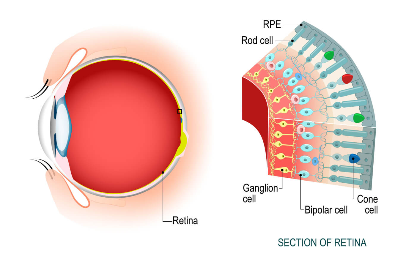
Unfortunately, the retina at the back of the eye does not have these elastic properties. This means it can’t spread incoming force to other areas and must bear the brunt of these injuries. This can affect cells in the retina called photoreceptors. They are responsible for turning light into nerve signals.
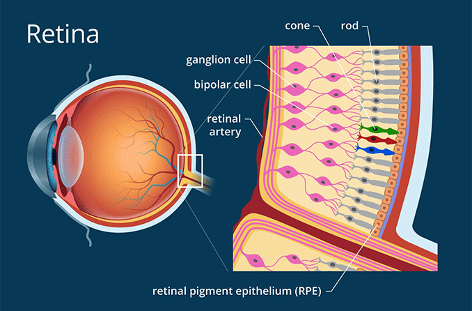
You have millions of color-sensitive cone cells in the back of each eye.
There are two types of photoreceptors: rods and cones. Cones, which process color, are responsible for sharp vision. Rods help you see in low light. After the rods and cones do their processing, the nerve signals are sent through the optic nerve to the brain. The brain then interprets the signals into the images we see.
Often, the photoreceptors that experience the most severe damage from the shock wave as it reaches the retina are those located in the macula (the center of the retina). The shock wave causes misalignment of the photoreceptors and swelling, which leads to the retinal cloudiness that characterizes commotio retinae. It is also possible for blood to leak into the retina and for pigment changes to occur.
Symptoms of commotio retinae
Commotio retinae is frequently asymptomatic, with no effect on a person’s vision. However, some individuals may experience:
Scotoma (blind spot)
Metamorphopsia (distorted vision)
A person may also experience symptoms resulting from the traumatic injury, including:
Hyphema – Blood pooling inside the anterior chamber of the eye. This is the section between the cornea (the clear layer at the front of the eye) and the iris.
Iridodialysis – When the iris separates from the sclera, the white of the eye.
Vitreous hemorrhage – Blood pooling in the vitreous cavity, the jelly-like substance inside the eye.
Choroidal rupture – A break in the choroid, a layer of blood vessels between the sclera and the retina.
Traumatic macular hole – A tear in the macula, the center of the retina that is responsible for central vision.
Orbital fracture – A break in the orbital bone (the eye socket).
Globe rupture – A break in the outer wall of the eye.
Posterior vitreous detachment – The gel in the eye (the vitreous) tears loose from the retina. This, in turn, can cause a retinal detachment.
Diagnosis of commotio retinae
If a patient has experienced ocular trauma, their eye doctor will typically perform a slit lamp exam to assess any damage. In this exam, they use a bright light, a special microscope and a hand-held lens to magnify and examine the internal structures of the eye. To get a better look at the retina, the doctor will likely perform a dilated fundus exam (also called ophthalmoscopy).
If they find signs of commotio retinae, the doctor may then use optical coherence tomography (OCT) to confirm their diagnosis. OCT is a non-invasive test that uses light waves to take pictures of the retina.
The patient rests their chin on a designated chin rest as the OCT machine scans the inside of the affected eye. The doctor then uses the resulting images to examine the different layers of the retina for damage. They can also refer to these images at future appointments to check for signs of healing.
Treatment options for commotio retinae
There is no official treatment for commotio retinae. Damaged photoreceptors often begin to heal starting around one week after initial injury. In many cases, a person will fully heal after around one month. However, in more serious cases, the patient may develop permanent damage.
In rare cases, some physicians may prescribe steroid injections, but this is not common practice.
How to protect your eyes from commotio retinae
Use of protective eyewear while playing sports is one of the best ways to protect your eyes from blunt trauma. This, in turn, protects your eyes from commotio retinae.
When to see an eye doctor
Seek immediate medical attention if you have experienced a traumatic injury. If you notice any changes in your vision after an injury, speak to your eye doctor.
Commotio retinae: A teaching case report. Optometric Education: The Journal of the Association of Schools and Colleges of Optometry. Winter-Spring 2023.
A brief history of dyslexia. The Psychologist. February 2018.
Commotio retinae. EyeRounds. February 2022.
Commotio retinae. EyeWiki. American Academy of Ophthalmology. April 2023.
Anatomy of the eye. Kellogg Eye Center. Accessed August 2023.
Retina. Cleveland Clinic. April 2022.
Unsafe at any speed. Review of Optometry. February 2021.
Hyphema. StatPearls [Internet]. December 2022.
Post-cataract surgery iridodialysis repair: A simple modified ab externo technique.Cureus. June 2022.
Anterior segment anatomy. American Academy of Ophthalmology. Accessed September 2023.
Vitreous hemorrhage. StatPearls [Internet]. July 2023.
Choroidal detachment. The Foundation of the American Society of Retina Specialists. Accessed September 2023.
Macula. Cleveland Clinic. May 2022.
Orbital fracture and traumatic injury. Dean McGee Eye Institute. Accessed September 2023.
Globe rupture. StatPearls [Internet]. December 2022.
What is optical coherence tomography? EyeSmart. American Academy of Ophthalmology. April 2023.
Page published on Tuesday, November 14, 2023
Page updated on Tuesday, November 21, 2023
Medically reviewed on Monday, November 6, 2023


