What your eye doctor can tell about your heart health
- How eyes can reveal potentially serious heart problems
- Eye conditions that reveal artery blockage, narrowing and hardening
- Eye conditions that reveal hypertension (high blood pressure)
- Heart disease
- Leaky aortic heart valve
- Can technology help eye doctors predict heart disease risk?
- The role of eye exams in detecting eye and heart disease

How eyes can reveal potentially serious heart problems
Your eye doctor can help keep an eye on your heart. Risk factors for heart disease like high blood pressure, diabetes and high cholesterol can be detected early during a comprehensive eye exam. In fact, your eye doctor may be the first to detect if you’re at risk for a heart attack or stroke.
Certain markers in the arteries and veins of the retina can provide eye doctors with important clues about what is happening in the small blood vessels of the brain and heart.
Eye conditions that reveal artery blockage, narrowing and hardening
Due to age and other risk factors such as high blood pressure and high cholesterol, arteries may narrow, harden and become clogged with deposits called “plaque.” This is known as atherosclerosis. It can affect blood vessels in the heart, brain and entire body, including small arteries supplying blood to the retina and optic nerve.
Small pieces of cholesterol plaque can sometimes break away from the carotid artery, a major blood vessel that branches off the aorta (from the heart) and supplies blood to the brain and eyes. These plaque deposits can travel to the arteries of the eye, making them visible during a dilated eye exam.
If these pieces of plaque reach the brain, they can cause a stroke. If your eye doctor spots plaque deposits, you will be sent to your primary care doctor, or to get an imaging study done.
Eye conditions that reveal artery blockage, narrowing and hardening include:
Amaurosis fugax
Amaurosis fugax can result from blockage of the internal carotid artery. It is a temporary loss of vision in one eye that can last up to 30 minutes. This is a warning sign that a stroke may happen.
Retinal artery occlusions
The most common cause of a retinal artery occlusion is an embolism, which is a blockage of an artery by a circulating blood clot. An embolism can travel from the heart or blood vessel and lodge in the eye. This can be visible during an eye exam.

Central retinal artery occlusion (CRAO)
The three most common types of blockages are due to cholesterol and calcium (from the carotid arteries that send blood from the heart to the brain) and blood clots from the valves of the heart. Your eye doctor can see the blockages as a white or orange blockage in your eye’s blood vessels.
A retinal artery occlusion is sometimes referred to as an “eye stroke” and can result in sudden, painless vision loss and difficulty seeing.
A 2015 study found that previously undiagnosed vascular (relating to blood vessels) risk factors were found in 78% of all central retinal artery occlusion patients. A 2017 study found that after a retinal artery occlusion, there was a high risk of a vascular event (such as a stroke) occurring within one month.
Retinal vein occlusions
Another type of “eye stroke,” a retinal vein occlusion (RVO) can indicate high cholesterol and high blood pressure. Retinal vein occlusions are the result of blood clots, narrow blood vessels or stiffened arteries that compress veins at junctions where they cross each other.

Sudden or gradual vision loss and blurry vision in part or all of one eye can result. An eye doctor may see signs of “arteriovenous nicking” — or constriction of the vein — where an artery crosses above it.
A 2016 South Korean National Health Insurance Service study analyzing over one million people found that the development of retinal vein occlusion was associated with a higher risk of a heart attack.
A 2019 meta-analysis delivered additional bad news. The analysis of 15 studies and nearly half a million people found that patients with RVO have an increased risk of cardiovascular events and death.
Xanthelasma
A possible sign of high cholesterol is the presence of yellow bumps or raised areas around the eyes and near the nose. These deposits, called xanthelasma, are the result of cholesterol that has built up under the skin.
While they do not affect your vision, they may indicate heart disease. Nearly half of individuals who have xanthelasma have high cholesterol.
Arcus senilis / Corneal arcus
This condition is caused by fatty substances and cholesterol that create a light gray or blue ring around the edge of the cornea.
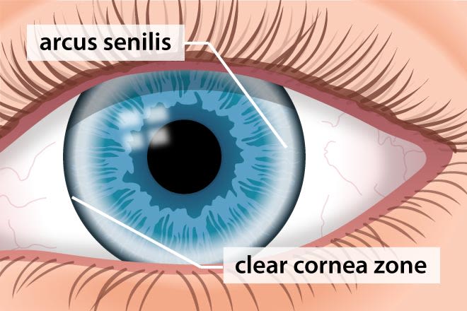
Appearance of arcus senilis (corneal arcus).
It is common in middle or older age, and is referred to as “arcus senilis” in this age group. But, it can be an indicator of high cholesterol in children or young adults (when it may be called juvenile corneal arcus).
Men younger than 40 with corneal arcus have an increased risk of death from coronary artery or cardiovascular disease compared with men who do not have it.
If an eye doctor finds the presence of corneal arcus in a patient younger than 40, a cholesterol screening with a primary care doctor will be recommended.
Eye conditions that reveal hypertension (high blood pressure)
It’s common for blood pressure to rise and fall during the day, but when blood pressure is consistently higher than normal, it’s called hypertension.
During a dilated eye exam, an eye doctor may suspect high blood pressure if they notice the following:
Damaged arteries in the eyes
Arteries in the eyes that are significantly smaller than the veins, or veins that are much bigger and bulging
Eye conditions that may indicate high blood pressure include:
Hypertensive optic neuropathy
Changes to the appearance of the optic nerve from high blood pressure are visible during a dilated eye exam. Optic neuropathy, or damage to the optic nerve, can result if blood flow to the optic nerve is blocked. Since the optic nerve carries signals from the retina to the brain, damage to its nerve fibers can cause vision loss.
Hypertensive retinopathy
Chronically high blood pressure can cause physical changes to blood vessels such as narrowing, “zig-zagging,” nicking and swelling. These changes are clearly visible during an eye exam. High blood pressure can eventually lead to bleeding in the eye, blurred vision or vision loss.
According to the NIH, there has been significant evidence that hypertensive retinopathy can be a predictor of disease or death due to organ damage from uncontrolled blood pressure.
Hypertensive chorioretinopathy/choroidopathy
Damage to the eyes’ blood vessels from high blood pressure can lead to the development of leaky blood vessels. This can lead to fluid building up underneath the retina (choroidopathy), which can be visible during a comprehensive eye exam. It can lead to distorted vision and retinal scarring.
Your eye doctor will recommend that you speak to a primary care doctor regarding high blood pressure if these conditions are detected during your eye exam.
SEE RELATED: High blood pressure: Hypertension's effect on eye health
Heart disease
Your eye doctor may be able to take an image of your eye to help detect heart disease earlier than ever before, according to the American Academy of Ophthalmology. Considering that heart disease is the leading cause of death in the world, diagnosing and treating heart disease early could help to prevent a heart attack or stroke.
Decreased blood flow due to heart disease can manifest in the retina, which receives nourishment from the central retinal artery. If the supply of blood to the retina is blocked or reduced, it is called “ischemia” and it results in retinal cell death.
When a retinal cell dies, it leaves a mark on the retina that is visible using technology called an OCT scan. By counting the number of marks left by dead retinal cells, eye doctors can detect whether a person may be at risk for a heart attack or stroke.
Leaky aortic heart valve
Aortic regurgitation is a condition that occurs when the heart's aortic valve does not close tightly. As a result, there is a large difference between a person’s systolic (when the heart beats) and diastolic (when the heart rests) blood pressure. Although it is rare, the pupils can dilate and constrict in sync with the heartbeat. This is referred to as Landolfi’s sign.
Can technology help eye doctors predict heart disease risk?
In 2018, Google’s health technology branch, Verily, published a study showing that an algorithm could use an individual’s retinal scans (current and historical) to predict their risk of cardiovascular disease.
The algorithm is programmed to identify which patients may suffer from a cardiac event in the next five years. The accuracy rate was 70%, similar to the blood tests that are currently used. The benefit is that this risk assessment can be done during an appointment at the doctor’s office, rather than having to wait for results from a blood lab.
The role of eye exams in detecting eye and heart disease
Comprehensive dilated eye exams play an important role in maintaining both eye health and heart health. In fact, an eye doctor may see markers for heart disease before any other symptoms are present. A comprehensive eye exam can detect life-threatening risk factors for heart disease and start a critical conversation about heart health.
The heart and the eye: Seeing the links. American Academy of Ophthalmology. December 2015.
8 Major health problems that an eye exam can detect. AARP. November 2021.
Amaurosis fugax. StatPearls [Internet]. September 2021.
Central retinal artery occlusion. StatPearls [Internet]. September 2021.
Carotid artery disease. Harvard Health Publishing. January 2015.
Cardiovascular risk factors in central retinal artery occlusion. Ophthalmology. September 2015.
Retinal artery occlusion and associated recurrent vascular risk with underlying etiologies. Plos One. June 2017.
High blood pressure, high cholesterol may be linked to retinal vein occlusion. American Academy of Ophthalmology. June 2008.
What can your eyes tell you about heart disease? UChicago Medicine. April 2018.
Retinal vein occlusion and the risk of acute myocardial infarction development: a 12-year nationwide cohort study. Scientific Reports. February 2016.
Association of retinal vein occlusion with cardiovascular events and mortality: A systematic review and meta-analysis. Retina. September 2019.
What is xanthelasma? American Academy of Ophthalmology. May 2021.
What Is arcus senilis? American Academy of Ophthalmology. April 2019.
Corneal arcus. Columbia Ophthalmology. Accessed 2021.
High blood pressure symptoms and causes. Centers for Disease Control and Prevention. May 2021.
High blood pressure and eye disease Information. Mount Sinai - New York. Accessed January 2022.
Hypertensive retinopathy. EyeWiki. American Academy of Ophthalmology. December 2021.
Hypertensive retinopathy StatPearls [Internet]. July 2021.
Early signs of heart disease appear in the eyes. American Academy of Ophthalmology. August 2021.
Retinal ischemia. Kellogg Eye Center University of Michigan. February 2015.
In your eyes: Clues to heart disease risk? Harvard Health. September 2021.
Prediction of cardiovascular risk factors from retinal fundus photographs via deep learning. Nature Biomedical Engineering. February 2018.
Page published on Wednesday, February 2, 2022


