Pyogenic granuloma: Causes, symptoms and prevention
What is a pyogenic granuloma?
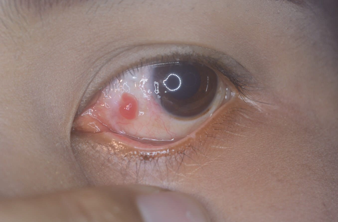
A pyogenic granuloma (PG) of the eyelid is a fleshy pink, red or purple growth on the conjunctiva — the lining that covers the inside of the eyelid and the white part of the eye. Eyelid PGs usually appear after someone has a stye, injury or surgery of the eye.
Pyogenic means “containing pus” and granuloma means “cluster of immune cells.” However, PGs do not contain either pus or clusters of immune cells, and the name that was given is considered inaccurate. Unfortunately, since the name has been around for a long time, everyone now uses it.
PGs can cover other parts of the body as well, most often on the inside of the mouth or nose and sometimes even on the skin. PGs of the eyelid are not dangerous and often go away without treatment.
When they don’t go away, there are a large number of effective treatments. Pyogenic granuloma of the eye and more serious eyelid lesions, such as cancer, may look the same. So, it is important to let a professional determine what you are dealing with.
Pyogenic granuloma of the eyelid
An eyelid pyogenic granuloma is a growth that contains many small blood vessels that each grow in small fingerlike projections called lobules. It usually grows on the part of the conjunctiva that is on the inside of the eyelid. But it can grow on the white part of the eye (sclera) and, rarely, on the cornea and skin of the eyelid.
Because PGs contain so many blood vessels, it is thought that an imbalance of blood vessel-forming growth factors is the underlying cause.
SEE RELATED: Chalazion: Symptoms, causes, treatment and prevention
Signs and symptoms
PGs appear as pink, red or purple fleshy growths that can be attached to the eye — usually on the inside of the eyelid — with or without a stalk. PGs grow rapidly (in weeks to months) to an average size of a quarter of an inch, which is about half the size of the iris (the colored part of the eye).
Because small PGs are often hidden on the inside of the eyelid, they may only be noticed when the eyelid is flipped inside out or the lesion grows too large to stay concealed. Large PGs can have an irregular surface that has tiny fingerlike (polypoidal) growths on the surface.
Because there are many small blood vessels inside a PG, it can easily bleed. An eyelid pyogenic granuloma is not painful, but it may cause symptoms of dry eye if it interferes with the normal blink action of the eyelid.
Causes and risk factors
The exact cause of PG is unknown. Since it is often associated with surgery and injury, it is thought to be a type of healing process.
Eye pyogenic granulomas are associated with:
Stye – In one study, an eye PG was preceded by a stye in 42% of all cases.
Eye or eyelid surgery – In the same study, surgery caused eye PG in 40% of the cases.
Eye injury – Injury caused eye PG in 5% of all cases in the study.
Human papillomavirus (HPV) infection – HPV can cause skin-tag-type growths on the surface of the eye. In some cases, it also causes an eye PG.
When a stye results in a pyogenic granuloma, it is generally because the stye has grown large enough to cause damage to the oil gland it grew inside of. Even though PGs can occur on the skin of the eyelid, studies showed that this is extremely rare. Almost all PGs occur on the inside of the eyelid.
PGs can form after eye surgeries, such as:
Draining of a stye
Eyelid “tuck” (blepharoplasty)
Surgery for an eye turn (strabismus)
Scleral buckle procedure for retinal detachment
Removal of growths from the surface of the eye
Surgery on the tear drainage (nasolacrimal) system
Injuries that are most likely to result in a pyogenic granuloma:
Blunt injury during sports
Motor vehicle accident
Work accident
Accidents from DIY or hobby projects
Other risk factors for developing a PG:
Age under 20 years old
Pregnancy or use of birth control pills
Port-wine stain (nevus flammeus) associated with Sturge-Weber Syndrome and surgery for these lesions
Eyelid inflammation (blepharitis)
Certain medications (retinoids for skin health, some HIV medications, cancer medications and immunosuppression medications)
Several of these risk factors, such as pregnancy and certain medications, have been studied in more detail for PGs that form in the mouth than for those that form in the eye.
Prevention
The most common causes of developing an eye PG are a stye and eye surgery. Of these two, there is not much one can do to avoid eye surgery. However, a lot can be done to avoid developing styes.
Styes are caused by plugging and inflammation of the oil glands of the eyelid, a condition called blepharitis. Blepharitis can be treated with regular application of warm compresses, cleaning of the eyelashes, eye drops, antibiotic pills and more.
READ MORE: Blepharitis: Causes, symptoms and treatment.
Treatment
Treatment can be as simple as doing nothing since many PGs will go away, given enough time. However, when a PG becomes large enough to interfere with blinking or becomes a cosmetic blemish, you may decide to treat it.
Since other growths on the eye that are riskier may look similar to a PG, including some cancers of the eye and eyelid, your doctor may decide that taking a biopsy to study the growth in more detail is the best first step.
Other treatment options can include:
Topical steroids – In one study, almost all (90%) of eye PGs went away when steroid eyedrops or ointments were used.
Steroid injection – A small needle is used to inject a steroid into the PG to make it shrink.
Bevacizumab injection – This drug, trade name Avastin, is used to treat swelling and new blood vessels growing in the retina but is also shown to shrink conjunctival PGs.
Surgical excision – This is the preferred method of removal when there is a suspicion of cancer and a biopsy is required.
Cryotherapy – This is when the growth is frozen off the eye. It is not used very often. It was shown to be effective when other treatments fail and the eye PGs keep coming back. A drug that slows cell division (mitomycin C) was used along with it.
Electrocautery – This procedure uses heat from an electric current to cut and remove tissue.
Laser ablation – In this process called photocoagulation, laser light is used to vaporize the PG, allowing precise blood vessel closure.
Timolol – This glaucoma medication is thought to shrink the blood vessels inside the PG and was shown to be an effective treatment that avoids the use of surgery or steroids.
Observation – Unless cancer is suspected, this is often the first thing to do as other treatments, such as steroids or surgery, can have undesirable side effects.
SEE RELATED: Relieving sore, painful eyelids
Complications
PGs can interfere with normal blinking, and this can result in dry eye and eye infections. The growths can rub open (ulcerate) and then become painful and infected. This is fortunately not common for eye PGs. The growths can bleed, especially when they become larger. While having blood coming out of your eye may be a scary experience, it is not dangerous.
Pyogenic granulomas, while they are actually very small growths, are noticed easily by other people, since they grow right on top of the eye. While PGs are not contagious, some people may not realize this and may be worried about getting infected. Other people may be distracted by the growth. Thus, cosmetic reasons are a common cause of surgical removal.
When to see an eye doctor
Only an eye doctor can tell for sure if you have a PG and provide the best treatment options and preventive strategies. It is important to remember that sometimes growths that look similar can be cancerous. If you have signs and symptoms of PG, see an optometrist or ophthalmologist to get an accurate diagnosis and receive the appropriate treatment.
Pyogenic granuloma. StatPearls. October 2022.
Pyogenic granuloma. EyeWiki. American Academy of Ophthalmology. July 2023.
Pyogenic granuloma. Cleveland Clinic. April 2022.
Conjunctival pyogenic granuloma during pregnancy. Arquivos Brasileiros de Oftalmologia. September 2021.
Sturge-weber syndrome. National Institute of Neurological Disorders and Stroke. January 2023.
Treatment of ocular pyogenic granuloma with topical timolol. JAMA Ophthalmology. April 2017.
What is a pyogenic granuloma? WebMD. April 2022.
Treatment of pyogenic granulomas with intralesional injections of bevacizumab. Investigative Ophthalmology & Visual Science. July 2019.
Keep it PG. Review of Optometry, December 2021.
Page published on Wednesday, August 2, 2023
Page updated on Tuesday, August 8, 2023


