Tests in an eye exam

Eye exam testing
A comprehensive eye exam includes tests designed to evaluate different parts of your eyes and vision. Eye doctors perform these tests to diagnose eye conditions, prescribe corrective lenses and determine treatment needs.
Routine eye exams are essential for both adults and children. They allow eye doctors to detect potential issues early — often before symptoms appear.
Certain tests in an eye exam can also show signs of systemic health conditions, such as diabetes and high blood pressure. Even if you have no noticeable eyesight issues, regular eye exams are important for your eyes, vision and general health.
Common tests in an eye exam
Eye exams include several basic tests that help eye doctors check your vision and screen for eye conditions. Some of the most commonly performed tests during an eye exam include:
Visual acuity test
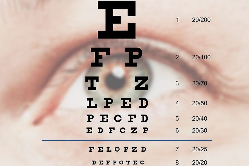
Visual acuity is a measurement of how clearly you see letters or objects at a specific distance. It is typically assessed using an eye chart, such as the Snellen chart.
A visual acuity test is usually conducted at a distance of 20 feet, with its results expressed as a fraction (such as 20/20). The top number represents your distance from the chart (20 feet). The bottom number indicates the distance that someone with “normal” vision can clearly see the same letters or objects that you see clearly at 20 feet.
Eye doctors use visual acuity tests to help determine if vision correction is needed. Visual acuity other than 20/20 could indicate a refractive error, which is a condition that affects how your eyes bend (refract) light for clear vision. It’s worth noting that eye strain can also indicate refractive error, even if a person has 20/20 vision.
READ MORE: What is 20/20 vision?
Refraction test
A refraction test helps determine the amount of vision correction needed for refractive errors, such as myopia (nearsightedness), hyperopia (farsightedness), astigmatism and presbyopia. Refraction may be performed manually or with an automated device.
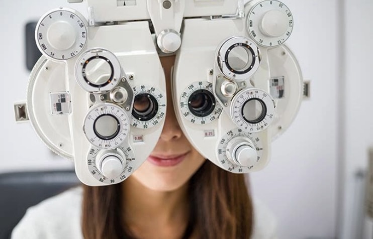
Manual refraction
During manual refraction, a phoropter — a device containing multiple lenses and dials — is placed in front of your eyes. As you look through different lenses, your doctor will ask you to read the letters from an eye chart. They will switch between lenses with different powers until you achieve your clearest vision. These results help determine your vision prescription.
Automated refraction
An autorefractor may also be used to perform a refraction test. You will look through a special machine as you focus on a specific target, such as an image. The machine will automatically measure your refraction to help determine your vision prescription.
Visual field test
A visual field test assesses how well you see objects in your peripheral (side) vision while looking directly ahead. Blind spots detected in your field of view may be an indication of glaucoma or other eye or brain problems associated with peripheral vision loss.
There are several types of visual field tests. One that is commonly used is the confrontation visual field test, in what is called the external examination (the pupils and eye muscles are tested, too).
Fluorescein eye stain test
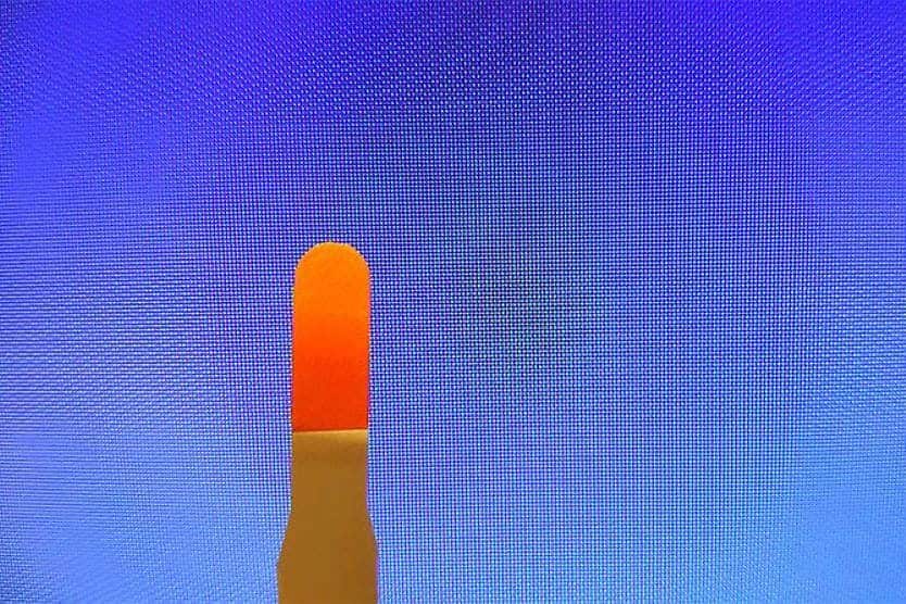
The fluorescein eye stain test involves placing an orange dye in the eye. The dye temporarily colors the tear film (the thin layer of tears covering the eye’s surface).
If you have a healthy cornea, the dye will remain on the eye’s surface until it’s washed away by your tears. But if you have any corneal damage, the dye can breach the surface and accumulate in deeper layers.
After the dye has settled, your doctor will shine a special blue light at your eye that causes any built-up dye beneath the surface to appear green. This helps your eye doctor to detect corneal scratches, blocked tear ducts and other issues.
Pupil dilation
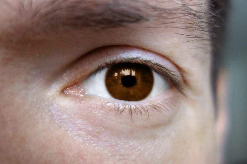
Before performing some eye exam tests, your eye doctor may dilate your pupils — the dark circles at the centers of your eyes — with special eye drops. This widens your pupils, allowing more light to enter and providing a clearer view of the back of your eye.
Slit-lamp exam
A slit lamp is a microscope that contains a focused light beam. Eye doctors use this instrument to perform a slit-lamp exam to check various parts of the eye. The slit lamp magnifies eye structures, allowing your doctor to see them in greater detail.
During this exam, you will rest your forehead and chin against the designated supports on the slit-lamp device. You will be asked to focus on a target while your eye doctor examines your eyes for potential issues.
Glaucoma tests
These tests help screen for glaucoma, a group of eye conditions that damage the optic nerve. While glaucoma does not have a definite cause, elevated eye pressure is a known risk factor. This increased intraocular pressure (or IOP) often results from fluid buildup in the eye.
Some glaucoma tests evaluate IOP, fluid drainage function and factors that can affect them. Other tests are used to detect optic nerve damage, peripheral vision issues and other signs of glaucoma. Glaucoma tests in an eye exam may include:
- Tonometry – This test uses a puff of air or a small instrument to measure eye pressure.
- Pachymetry – This test allows doctors to gauge corneal thickness (the cornea is the clear front layer of the eye). The thickness of the cornea may affect the tonometer’s accuracy in measuring IOP.
- Gonioscopy – With this test, your doctor can examine the angle where the cornea and iris (the colored part of the eye) meet. This helps determine if fluid is draining properly from the eye.
Special glaucoma imaging tests may also be used to further evaluate the optic nerve.
SEE RELATED: What is a glaucoma suspect?
Color blindness test
Color blindness tests help detect color vision deficiencies, or the inability to see or distinguish between certain colors. Eye doctors may use various color blind tests during an eye exam.
The most common color blind test is the color plate or Ishihara test for colour deficiency. It features a series of “plates” or charts containing patterns of colored dots forming symbols or numbers. People with normal color vision will see specific figures. Those with color vision deficiencies may perceive them differently or not at all.
SEE RELATED: What are the different types of color blindness?
Extraocular motility (EOM) assessment
An EOM assessment evaluates your ability to move your eyes in different directions. It can help detect problems with eye muscle movement.
For this test, your eye doctor will hold a finger or an object (such as a pen) in your direct line of vision. While keeping your head still, you’ll be asked to follow the object with your eyes as it’s moved in various directions.
Stereopsis (depth perception) test
A stereopsis test measures depth perception, which is the ability to see objects in three dimensions (3D) and accurately judge distances.
Your eye doctor may use different methods to test your depth perception, including:
- Viewing devices – You’ll look into a special device and each eye will be shown a different image.
- Special glasses – You’ll view a picture or an object while wearing polarized 3D glasses.
- Physical objects – You’ll observe or touch physical objects placed at various distances away from you.
Your responses during these tests help determine how well you perceive depth and judge distance.
Cover test
Cover testing can help check for signs of eye misalignment (strabismus). For a “cover-uncover” test, you’ll be asked to look straight ahead at a distant target. One of your eyes will be covered for a few seconds. As the cover is removed, your doctor will observe whether the direction of your gaze shifts, which could indicate misalignment. The process will then be repeated on your other eye.
In another version, called an “alternate cover test,” your eye doctor may alternate covering each eye to check for any gaze shifts in the eye that is not covered.
Specialized eye tests in an eye exam
A comprehensive eye exam may include specialized tests. These tests allow for a more detailed evaluation of individual eye structures. They can include:
Retinoscopy
Retinoscopy helps your doctor detect and evaluate refractive errors by assessing how light reflects off the retina. To perform this test, your eye doctor will use a handheld device to direct a beam of light toward the retina at the back of your eye.
Retinoscopy helps your doctor determine if a refractive error exists and the level of vision correction and prescription needed to help you achieve clear vision.
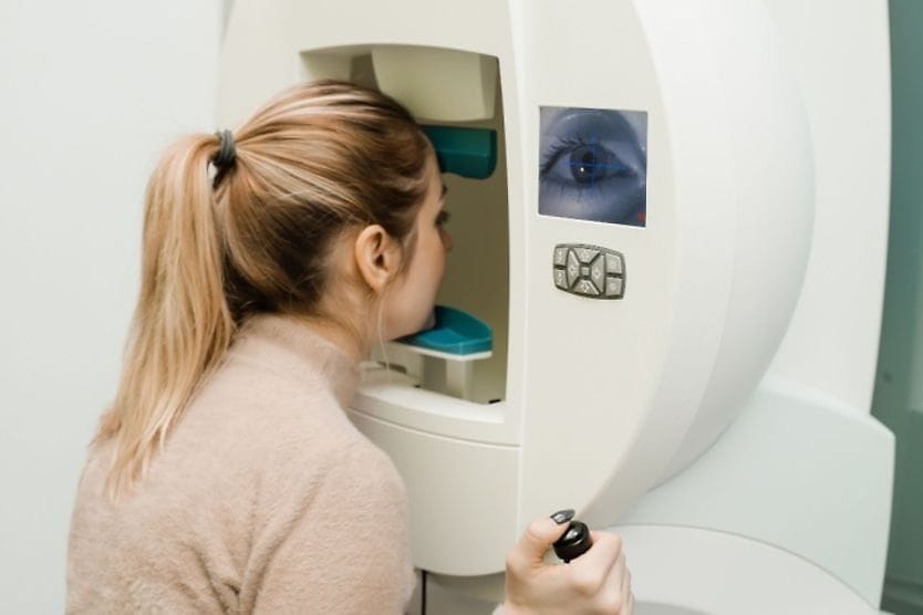
Corneal topography
Corneal topography is an imaging technique used to create a map of the cornea’s surface. For this exam, you will be positioned in front of a bowl-shaped device featuring a series of lighted circles. After placing your forehead and chin against the designated supports, you will look at a specific target while the device scans your eyes.
Eye doctors use these scans to detect corneal damage, disease and irregularities.
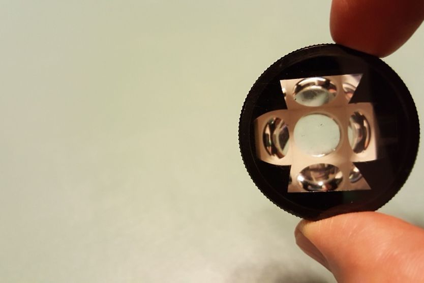
Ophthalmoscopy (fundoscopy)
Ophthalmoscopy allows eye doctors to see structures in the fundus (the back of the eye). This test aids in screening for conditions like retinal detachment and glaucoma. It may be performed using one of three techniques:
- Direct ophthalmoscopy – A handheld device containing a bright light and a series of lenses are used to see the back of the eye.
- Indirect ophthalmoscopy – A head-mounted light and special lens placed in front of the eye are used for viewing the fundus.
- Slit-lamp ophthalmoscopy – A slit-lamp microscope and a small lens placed in front of the eye are used to see the fundus.
Your pupils may be dilated for this exam depending on the technique used.
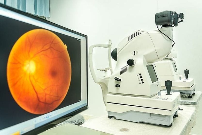
Optical coherence tomography (OCT)
Optical coherence tomography is an imaging test that takes pictures of the retinal layers using light waves. Eye doctors use these images to:
- Assess the thickness of the retina
- Diagnose and monitor retinal diseases
- Evaluate conditions affecting the optic nerve (such as glaucoma)
Your doctor may dilate your pupils before beginning the OCT scan. Once positioned in front of the device, you will be asked to rest your forehead against the designated support as the scanner captures images of your retina or optic nerve.
Contact lens fittings
A contact lens exam and fitting help your eye doctor assess whether contact lenses are right for your vision needs. This process involves taking specific eye measurements to ensure the best lens type and fit.
As part of the exam, your eye doctor will:
- Measure the shape and curvature of your cornea
- Recommend the ideal type and brand of contacts for your eyes
- Provide you with trial contact lenses to assess their fit
A contact lens exam and fitting are separate from the routine eye exam and typically require extra time and an additional fee. If you wear contact lenses or want to start, let your doctor’s office know when scheduling your eye exam.
SEE RELATED: Are contact lens and eyeglass prescriptions the same?
Vision screening vs. comprehensive eye exam
Vision screenings help detect potential vision issues and determine if further evaluation is necessary. Unlike a comprehensive eye exam and testing from a licensed eye care provider, vision screenings do not assess your overall eye health, provide a diagnosis or prescribe glasses.
Screenings are commonly performed:
- In schools
- During medical checkups
- As part of driver’s license renewals
- During sports assessments
They typically measure distance vision and may include basic color vision and eye movement tests as well.
While they can provide helpful information, vision screenings are not a substitute for a comprehensive eye exam. It’s important to schedule a full eye exam with your eye doctor each year. This gives them a chance to detect and diagnose eye conditions, update your vision prescription, determine treatment needs, and support long-term eye health.
READ NEXT: What’s the difference between an optometrist and an ophthalmologist?